
Vascular Ultrasound Systems and Doppler in Cardiology
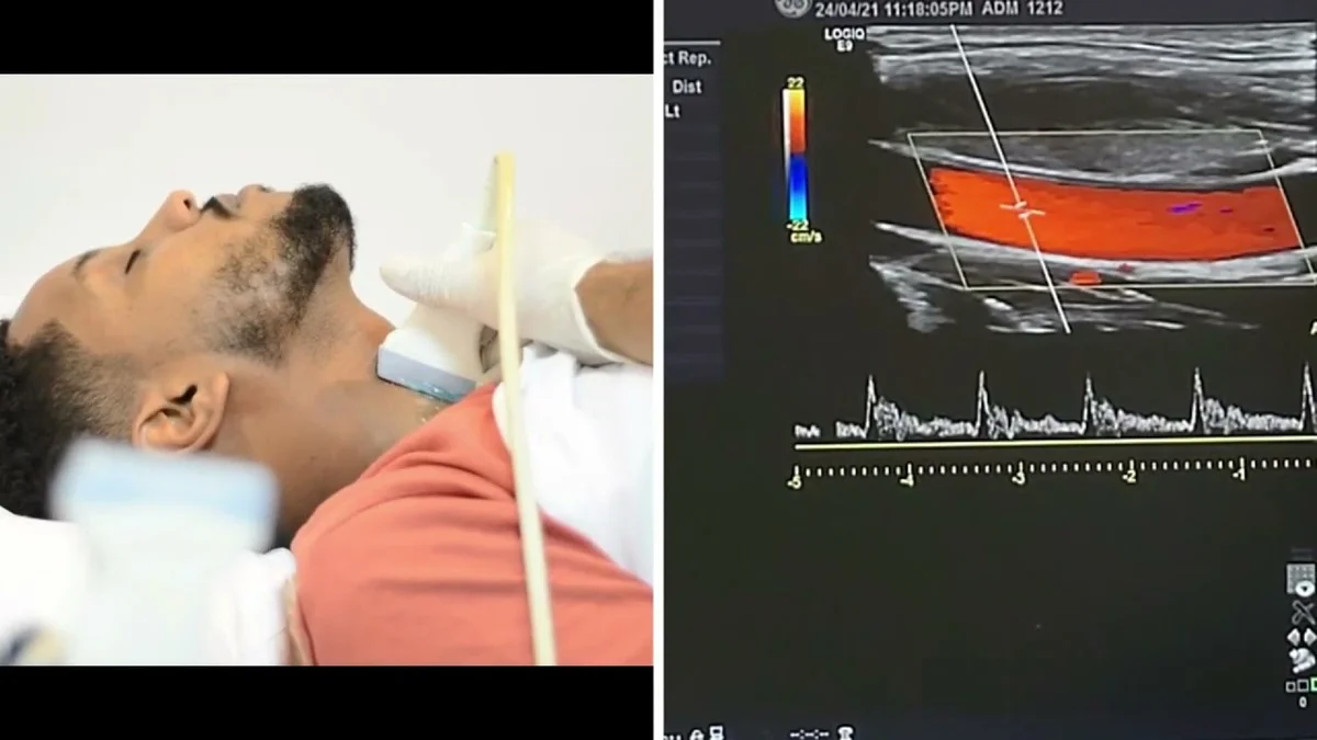
Vascular ultrasound is a non-invasive imaging method for assessing the morphology and hemodynamics of arteries and veins. Duplex and triplex scanning combine B-mode with Doppler assessment of blood flow velocity.
Vascular Ultrasound Methods
Modern vascular protocols include:
- Duplex scanning: B-mode + color Doppler, visualizing the vessel wall and flow;
- Triplex scanning: B-mode + color Doppler + spectral Doppler, quantifying velocity;
- Power Doppler: increased sensitivity to slow flow, without directional information;
- B-Flow (GE): display of blood flow without Doppler artifacts, at high resolution.
Criteria for Assessing Carotid Stenosis
Ultrasound criteria for stenosis grading according to NASCET:
| Degree of stenosis | PSV, cm/s | ICA/CCA ratio |
|---|---|---|
| < 50% | < 125 | < 2.0 |
| 50–69% | 125–230 | 2.0–4.0 |
| ≥ 70% | > 230 | > 4.0 |
| Occlusion | No flow | — |
Choosing an Ultrasound System for a Vascular Laboratory
Key features of a system for vascular examinations:
- A 5–12 MHz linear transducer for superficial vessels (carotid, peripheral);
- A 2–5 MHz convex transducer for the abdominal aorta and iliac arteries;
- Automatic calculation of the ABI (ankle-brachial index);
- Automatic measurement of IMT (intima-media thickness);
- Elastography software for characterizing plaque;
- Integration with PACS and the cardiology information system.
Conclusion
A vascular ultrasound system is one of the most sought-after investments for a cardiology clinic. KombiMED selects the optimal configuration based on the clinic's profile, examination volume and budget.
Equipment
Equipment for this topic
Vascular Ultrasound Systems
Ultrasound systems for vascular diagnostics: Doppler, ABI, carotid scanning
Related articles
Planning a purchase in this area?
Tell us what your laboratory needs and we will send you a tailored offer.
Contact KombiMED