
Digital Pathology Systems: A New Era in Morphological Diagnostics
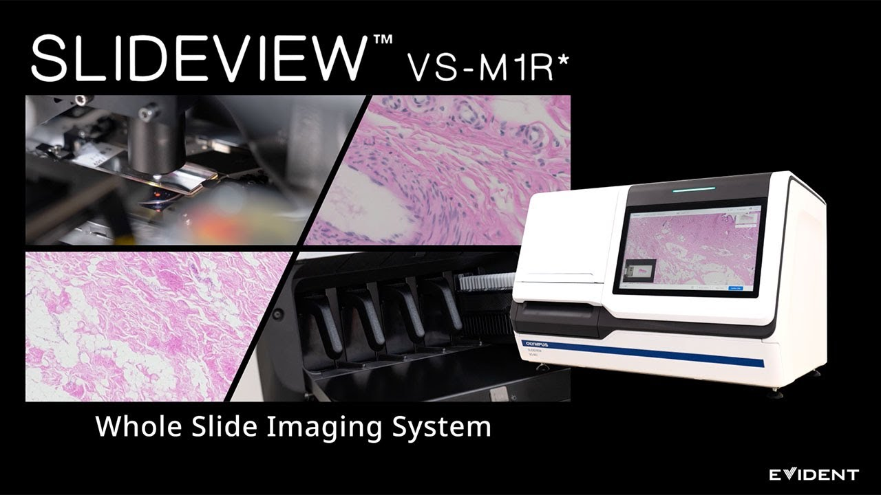
Digital pathology is transforming classical morphological diagnostics, moving from manual slide review to high-speed scanning, centralized storage and remote interpretation of Whole Slide Images (WSI). KombiMED supplies comprehensive solutions for a laboratory's transition to digital workflows.
What Is Digital Pathology?
Digital pathology is a field of medical informatics in which histological and cytological slides are digitized with high-speed slide scanners and analyzed on a monitor screen instead of through a microscope eyepiece. The technology is based on Whole Slide Imaging (WSI): creating a full-resolution digital image of the entire glass slide at a resolution of up to 0.25 µm/pixel.
Unlike the traditional approach, digital systems provide:
- A single data repository: all images are archived in a centralized system with instant access.
- Multi-vendor compatibility: support for scanners from different manufacturers within one platform.
- Remote diagnostics: the ability to consult on and verify a diagnosis without physically moving slides.
- AI integration: connection of artificial intelligence modules for automated screening and quantitative analysis.
Key Components of a Digital Pathology System
Slide Scanners
High-throughput scanners create WSI at 20× or 40× magnification in 60–120 seconds per slide. Modern models support continuous loading of up to 400 slides and operation in brightfield, fluorescence and multispectral imaging modes.
Example solutions: Leica Aperio GT 450 DX, Hamamatsu NanoZoomer S360MD, 3DHISTECH PANNORAMIC 1000, Philips IntelliSite Ultra Fast Scanner.
Image Management Systems (IMS)
The Image Management System is the "heart" of digital pathology. The IMS receives images from scanners, indexes them, links them to the patient case, and handles versioning and access control.
Example: PathoZoom® Digital Lab by Smart In Media, a comprehensive digital pathology platform with an integrated IMS and support for AI modules, remote diagnostics and multi-vendor scanners. The system is CE-certified and supports the DICOM and HL7 standards.
Visualization and Viewing Tools
Specialized viewers that support navigation through gigapixel images, annotation, side-by-side comparison of multiple slides, measurements and simultaneous collaboration among several pathologists.
Benefits of Digital Pathology
Instant access to archived and current cases shortens the time from biopsy receipt to final report.
Calibrated scanners and digital protocols eliminate the subjectivity associated with microscope and illumination quality.
Digital archives with backups protect against the loss of unique slides.
The platform grows with the laboratory, from a single scanner to full automation.
Integration with the LIS, HIS and PACS creates a single digital pathway from test order to final report.
Digital slides are used for resident training, multidisciplinary tumor boards and clinical research.
- Faster diagnosis
- Standardized quality
- Data security
- Scalability
- Workflow continuity
- Education and research
Applications
Histology
Digital histology covers the full range of stains: H&E, immunohistochemistry (IHC), fluorescence in situ hybridization (FISH) and special stains. WSI images become the basis for quantitative biomarker analysis and companion diagnostics. Automated HER2 status determination, Ki-67 counting and PD-L1 expression scoring are performed with a precision that manual counting cannot achieve.
Cytology
Liquid-based cytology (ThinPrep, SurePath) is integrated with scanners and AI modules for automated Pap test screening, which increases sensitivity to up to 95% and reduces the cytologist's workload. Cytology solutions include the Landing AI Cytology and Hologic Genius Digital Diagnostics platforms, which cover the full cycle of digital cytological analysis.
Molecular Pathology
Digital platforms combine morphological data with the results of molecular testing (NGS, PCR) to create multimodal reports for oncology tumor boards. Integrating Whole Slide Imaging data with genomics, transcriptomics and proteomics opens the way to multi-omics diagnostics, a comprehensive approach that makes it possible to predict response to therapy based on the combined data.
Early Cancer Detection
AI integration with digital pathology enables early detection of cancer through analysis of WSI slides, biochemical data and companion diagnostics (CDx). Tempus Digital Pathology, Paige Alba™ and Evident Digital Pathology solutions process scans, images and clinical data to generate digital biomarkers and predict response to targeted therapy.
Integration into the Laboratory Infrastructure
Digital pathology does not exist in isolation. Integration with the following systems is critical:
Integration protocols: HL7 v2/FHIR, DICOM, IHE Anatomic Pathology Workflow.
- LIS (Laboratory Information System): Dedalus GLIMS, LabWare LIMS, CGM LAB, Epic Beaker AP, 1C LIS
- HIS (Hospital Information System): Dedalus ORBIS, Epic, Oracle Cerner, SAP IS-H, OpenMRS
- PACS/DICOM: standardized storage and transfer of images on par with radiology data
KombiMED Solutions
KombiMED provides complete equipment for digital pathology laboratories:
Request a consultation: our specialists will help you design the optimal solution based on your laboratory's sample volume, tasks and budget.
*KombiMED: centralized procurement of medical equipment from Europe. Deliveries to Kazakhstan, Central Asia and the Caucasus with full-cycle support, from design to staff training.*
- Selection and supply of slide scanners from leading manufacturers
- Implementation of image management platforms (PathoZoom® Digital Lab)
- Integration with existing LIS/HIS
- Staff training and technical support
- IT infrastructure design: servers, storage, network
Related articles
AI & Software
AI & Software
AI & Software
Planning a purchase in this area?
Tell us what your laboratory needs and we will send you a tailored offer.
Contact KombiMED