
PACS/DICOM Integration for CT, MRI and Mammography
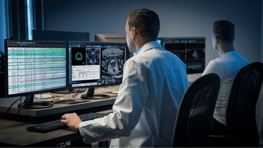
PACS and DICOM are the foundation of digital diagnostic imaging. A unified picture archiving and communication system brings CT, MRI, mammography, X-ray and digital pathology data together in one ecosystem. KombiMED designs and implements PACS infrastructure for institutions of any size.
What Are PACS and DICOM?
PACS (Picture Archiving and Communication System) is a system for archiving and transmitting medical images that handles the receipt, storage, display and distribution of images from different modalities.
DICOM (Digital Imaging and Communications in Medicine) is the international standard for transmitting and storing medical images. DICOM defines the file format, network protocols and display specifications, ensuring interoperability between equipment from different manufacturers.
Together, PACS and DICOM form the basis for:
- Filmless radiology, with film archives eliminated entirely
- Instant access to images from anywhere in the institution
- Integration with the HIS and LIS to build a complete medical record
- Teleradiology and remote consultations
PACS for the Main Modalities
Computed Tomography (CT)
CT generates the largest volume of data in the radiology department. A single multislice study can contain 500–3,000 slices.
PACS requirements:
- High-speed data ingestion (at least 1 Gbit/s)
- Support for 3D reconstruction, MPR, MIP, VRT
- AI integration for automated pathology detection
- Compatibility with leading CT systems: Siemens NAEOTOM Alpha, GE Revolution CT, Philips Spectral CT
Magnetic Resonance Imaging (MRI)
MRI requires storing multiple sequences (T1, T2, FLAIR, DWI, contrast enhancement) for each study. A single study can be up to 2 GB.
PACS requirements:
- Support for multi-series studies with correct grouping
- Fusion imaging: overlaying MRI on CT or PET
- Advanced measurement tools for neuroradiology
- Compatibility: Siemens MAGNETOM Vida, GE SIGNA Premier, Philips Ingenia Ambition
Mammography
Digital mammography, including tomosynthesis (3D mammography), generates high-resolution images with strict display accuracy requirements.
PACS requirements:
- Support for mammography viewers with 5-megapixel monitors
- Display of tomosynthesis as a slice series and in C-View mode
- AI assistance for CAD (Computer-Aided Detection)
- Compatibility: Hologic 3Dimensions, Siemens MAMMOMAT Revelation, GE Senographe Pristina
PACS Architecture
System Components
- DICOM server: central image repository indexed by patient, study, series and instance
- Workstations: specialized diagnostic terminals with calibrated monitors
- Web viewer: browser-based image access for clinicians without diagnostic workstations
- Router: automatic distribution of images to worklists based on modality, urgency and specialty
- Archive: long-term storage with a tiered strategy: online (SSD), near-line (NAS), archive (tape/cloud)
Integration with Information Systems
- RIS (Radiology Information System): management of schedules, protocols and reports
- HIS (hospital information system): a single medical record with image access from any module
- LIS: correlation of radiology and laboratory data
- Digital pathology: DICOM WSI is integrated into the shared archive on an equal footing with radiology images
PACS and Digital Pathology: Convergence
The current DICOM standard (Supplement 145) defines a format for whole slide images, which makes it possible to store pathology images in the same PACS as CT, MRI and mammography. This provides:
- A single point of access for all image types
- Correlation of radiology findings with histological confirmation
- Multidisciplinary tumor boards with simultaneous review of all modalities
- Unified backup and disaster recovery
Benefits of a Unified PACS Infrastructure
Images are available within seconds from any authorized terminal. Physicians no longer wait for films or discs to be transferred.
Consolidated storage instead of multiple isolated systems reduces total cost of ownership by 30–40%.
Centralized access management, encryption and an audit trail. Compliance with GDPR and ISO 27001.
From a single department to a multi-site network with a cloud archive, the PACS grows with the institution.
Remote radiologists access studies via a secure VPN or a cloud portal.
- Speed of access
- Storage savings
- Data security
- Scalability
- Teleradiology
Leading PACS Solutions
- Sectra PACS: multimodal platform supporting radiology, cardiology and pathology
- Agfa HealthCare Enterprise Imaging: a single platform for all types of medical images
- Philips Vue PACS: cloud architecture with AI integration
- GE Centricity PACS: a solution for large radiology departments
- Horos / OsiriX: open DICOM viewers for macOS with plugin support
KombiMED Solutions
KombiMED designs and implements end-to-end PACS infrastructure:
Request a consultation: our engineers will design a PACS architecture tailored to the specific needs and scale of your institution.
*KombiMED: centralized procurement of medical equipment from Europe.*
- Audit of the current IT infrastructure and assessment of data volumes
- Design of a PACS architecture covering all modalities
- Supply of server hardware and workstations
- Integration with CT and MRI scanners, mammography systems and WSI scanners
- Configuration of interfaces with the HIS, RIS and LIS
- Migration of archives from existing systems
- Staff training and technical support
Related articles
AI & Software
AI & Software
Planning a purchase in this area?
Tell us what your laboratory needs and we will send you a tailored offer.
Contact KombiMED